
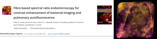
https://doi.org/10.1364/BOE.10.001856
I really like this one, tackling a Proteus challenge of making those annoying green fluorophores stand out against the green lung tissue in fluorescence imaging. But green doesn’t just mean green, it’s all about the shades of green…..
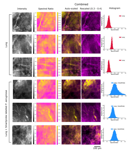
So, in fluorescence imaging when pumped with blue light, normally the resulting “green” light coming back is all dumped into one channel. But actually, different objects have different spectra and can be distinguished.
But if you don’t want to do full (expensive or slow) hyperspectral imaging, we came up with an alternative for fibre based imaging.
Just splitting the green channel in two, and using this to create a fluorescence “Spectral ratio”, all those greens can be made to look very different.
Key is doing this per core of the fibre:
– avoiding chromatic abberation challenges of spectral imaging
– making this applicable to fibred imaging
– overcoming the different spectral modes in the fibre cores which would make using standard spectral imaging techniques fail.
All thanks to the hard work of my student Helen – nice work!

One Reply to “”